XPS imaging is useful in understanding:
- Distribution of surface chemistry across a surface
- Determination of the limits of contamination
- Examination of thickness variation of ultra-thin coatings.
Two approaches for obtaining XPS images: mapping (serial acquisition) or parallel imaging (parallel acquisition).
Mapping
Serial acquisition of images is based on a two-dimensional, rectangular array of small-area XPS analyses. This method enables measurements of the distribution of elements or chemical states. Serial acquisition is generally slower than parallel acquisition but can collect a range of energies at each pixel compared with collecting only a single energy for parallel acquisition. Using this method, the analysis position is fixed and the specimen surface is moved with respect to this position.
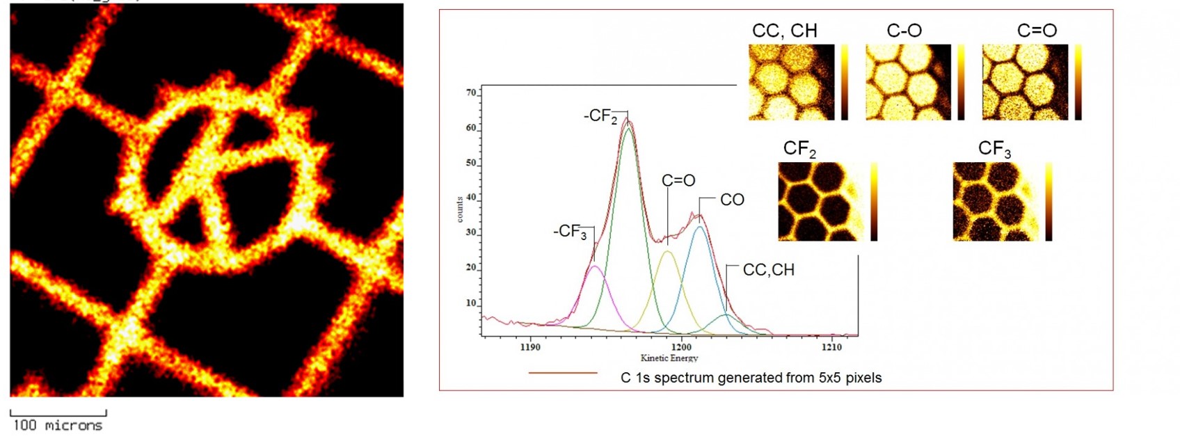
Parallel imaging
Parallel acquisition method simultaneously images the entire field of view without scanning voltages applied to any spectrometer component. This parallel imaging provides that a set of quantitative images can be acquired in only a few minutes. This method of imaging is faster and produces an better image resolution, of <3 μm, than the mapping method. Then subsequent data processing produces the relative atomic concentration image that can be used to define the elemental and chemical composition as a function of position.
SnapMap imaging
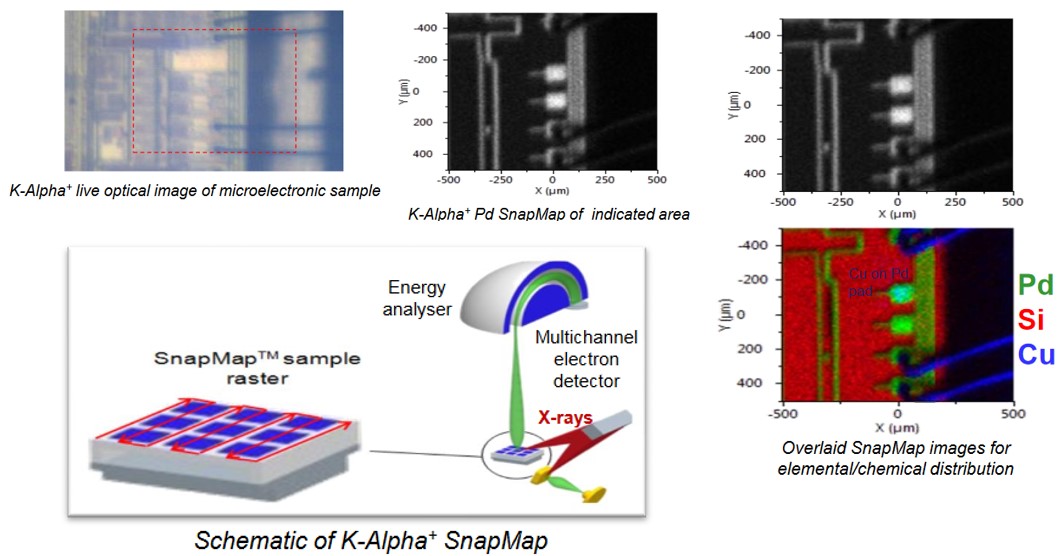
“SnapMap” imaging allows for rapid aquisition of image data of a surface.
In such “snapmap” images, XPS spectra are acquired on-the-fly as the sample is rastered on the stage without the stage needing stop at each analysis point.
Regions sizes from 0.5mm x 0.5mm up to 3mm x 3mm can be mapped at a resolution down to 30µm, whilst larger regions being mapped by “stitching” smaller areas as common with large scale XPS imaging. This technique has already been used to help identify corrosion distribution for a multi-national company.