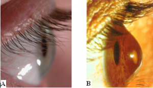
Keratoconus (literally “cone-shaped cornea”) is a non-inflammatory corneal dystrophy in which the normally dome-shaped cornea bulges outward. The condition, normally presents in the teenage years as irregular astigmatism, with a blurring of vision or glare and streaking of lights at night. As the condition progresses, the cornea gradually becomes thinner and weaker, and secondary complications may occur such as tearing and scarring within the cornea. In Europe, keratoconus is estimated to occur in about 1 in 375 of the general population [1] but its incidence is known to vary world-wide.
Although the precise cause and underlying pathological mechanism of keratoconus is unknown, both environmental and genetic factors are thought to contribute to its development [2]. Collagen is known to play a major role in maintaining the strength and shape of the healthy cornea but in keratoconus, it is thought that an upregulation of enzymes leads to the degradation and slippage of the collagen which in turn, leads to further corneal thinning and shape change [3].
[1] Godefrooij DA, et al. Age-specific incidence and prevlance of keratoconus: a nationwide registration study. Am J Ophthalmol 2017; 175: 169-172
[2] Davidson AE, et al. The pathogenesis of keratoconus. Eye. 2014; 28: 189-195.
[3] Meek KM, et al. Changes in collagen orientation and distribution in keratoconus corneas. Invest Ophthalmol Vis Sci. 2005; 46: 1948-1956.

